Orthopedic care is entering a period of measured expansion. The World Health Organization reports that approximately 1.71 billion people live with musculoskeletal conditions worldwide. These conditions affect mobility, work, independence, and daily comfort. Behind every statistic is a patient choosing between a painful staircase and a safer walk.
The market is also becoming more specialized. Grand View Research estimates that the global orthopedic devices market was worth about USD 48 billion in 2023, with continued growth expected through 2030. MarketsandMarkets identifies joint reconstruction, spinal devices, trauma fixation, and orthopedic accessories as important market segments. These reports use different definitions, so their figures should not be compared without care. Forecasts are useful, but they are not flawless.
This 2026 guide examines the major types of orthopedic implants and instruments used in contemporary clinical practice. It considers joint replacement systems, trauma plates, intramedullary nails, spinal implants, arthroscopy tools, surgical navigation, and supporting instruments. Materials such as titanium alloys, cobalt-chromium, ceramics, and advanced polymers receive practical attention. Design matters. So does handling. A precisely engineered implant can still underperform when sizing, sterilization, surgical technique, or patient selection is overlooked.
Clinical evidence remains central. The American Academy of Orthopaedic Surgeons emphasizes appropriate indications, patient safety, and evidence-based decision-making. Manufacturers must also support traceability, quality systems, and transparent performance data. This overview does not treat every new device as a breakthrough. Some innovations deserve enthusiasm; others require longer follow-up. That distinction protects surgeons, hospitals, and patients while encouraging better orthopedic care.
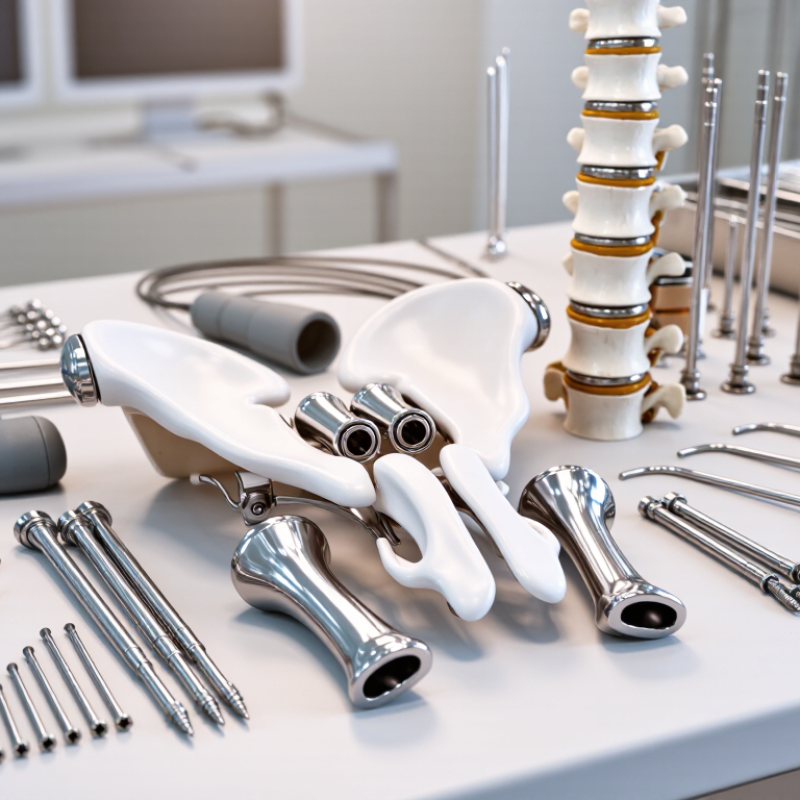
Orthopedic implants are medical devices placed inside the body to support, replace, or stabilize damaged bones and joints. Plates, screws, nails, and fixation wires help maintain alignment during healing. Joint implants replace worn surfaces in hips, knees, or shoulders. Their clinical role depends on anatomy, injury severity, bone quality, and patient activity.
Orthopedic instruments are the tools used to prepare tissue and position implants accurately. Drills create controlled holes, while guides help surgeons maintain proper angles. Retractors improve visibility. Trial components allow assessment before final placement. In practice, experienced teams select instruments that match the implant system, surgical approach, and patient-specific anatomy. Small errors can affect stability, movement, or recovery. That deserves honest attention.
Tips: Confirm implant measurements before preparation. Check instrument function and sterility according to approved protocols. Use imaging when alignment is difficult to judge. Document implant size, location, and intraoperative findings clearly. Never treat a standard instrument set as universally suitable. Bone density varies. Patient anatomy varies more.
Clinical outcomes also depend on surgical technique, rehabilitation, infection prevention, and follow-up. No implant removes every risk. A well-designed device may still perform poorly if positioning is inaccurate or healing is limited. Continuous review of surgical results can reveal patterns that are easy to miss during a busy procedure.
Definitions and Clinical Roles
The chart compares major orthopedic implant families by the number of representative device examples included in each category. Joint replacement devices restore damaged articulating surfaces, fracture-fixation systems stabilize broken bones, spinal implants support alignment and fusion, sports-medicine implants secure soft tissues, and bone-graft substitutes help fill defects or support bone healing.
Orthopedic implants are selected according to anatomy, load, bone quality, and expected activity. The major categories include joint replacements, fracture fixation devices, spinal implants, and sports medicine anchors. Joint replacements restore damaged hips, knees, shoulders, or smaller joints. They often combine metal or ceramic components with low-friction bearing surfaces. Fit matters. A small mismatch can affect movement, stability, or wear.
Fracture fixation implants include plates, screws, intramedullary nails, wires, and external fixation systems. Surgeons choose them according to fracture pattern, soft-tissue condition, and healing goals. Spinal implants may include cages, rods, screws, and motion-preserving devices. Their placement requires careful imaging and knowledge of nerves, alignment, and biomechanics. Sports medicine implants often use suture anchors or interference screws for tendon and ligament repair. Material choice can influence strength, tissue response, and imaging visibility.
Orthopedic instruments support every stage of treatment. Cutting guides, drills, reamers, trial components, forceps, and reduction tools must match the procedure. In practice, instrument handling can affect accuracy as much as implant design. Sterility, calibration, traceability, and maintenance deserve equal attention. Even careful planning may miss an anatomical variation. That possibility requires cautious imaging review and sound clinical judgment. Patient-specific evidence should guide the final selection.
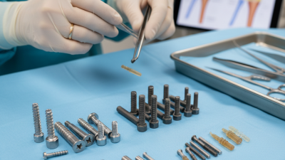
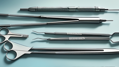
Orthopedic surgical instruments are the working core of implant procedures. Common types include bone saws, drills, reamers, osteotomes, rongeurs, retractors, graspers, and depth gauges. Each tool controls a specific action: cutting, shaping, holding, or measuring bone. A drill with adjustable speed can reduce heat during preparation. A sharp osteotome creates controlled cuts, while a stable retractor protects soft tissue from unintended pressure.
Instrument choice should follow the procedure, anatomy, and bone quality. Trauma cases often require reduction forceps, guidewires, depth gauges, and torque-limiting drivers. Joint reconstruction usually adds cutting blocks, rasps, trial instruments, and alignment guides. Minimally invasive techniques may depend on slim retractors and angled instruments. The World Health Organization reports that about 1.71 billion people live with musculoskeletal conditions worldwide (WHO, 2022). That burden increases demand for reliable surgical systems, but demand alone should not dictate tray design.
Sterilization and traceability matter as much as mechanical precision. AAMI ST79 emphasizes validated cleaning, inspection, packaging, and sterilization practices for reusable medical devices. Instruments with blocked channels or worn jaws can compromise workflow. A practical caveat remains: no tray is perfect. Excess instruments increase handling and processing time. Too few instruments create delays. The better approach combines surgeon feedback, maintenance records, and procedure-specific data. Industry market analyses also forecast continued growth in orthopedic devices through 2030, although estimates vary by methodology (Grand View Research, 2024). That variation deserves attention before hospitals make purchasing decisions.
Orthopedic implants must match both anatomy and patient needs.
Material choice affects strength, weight, corrosion resistance, and tissue response. Titanium alloys are valued for their lower weight and bone-friendly stiffness. Cobalt-chromium alloys provide strong wear resistance in demanding joint applications. Stainless steel remains useful for selected fixation devices and temporary support.
Design details matter as much as material. A plate may need a low profile, rounded edges, and variable-angle screw holes. These features can reduce soft-tissue irritation and improve fixation around complex fractures. Joint components require smooth bearing surfaces, accurate geometry, and controlled motion. Small changes in taper fit or surface texture can influence stability. Very small differences matter.
Compatibility requires careful review. Surgeons and engineers should confirm dimensions, screw threads, instruments, imaging requirements, and sterilization limits before use. Mixing components from different systems may create poor fits or unexpected mechanical behavior. Patient bone quality also changes the decision. A strong implant can still fail when fixation is weak.
No material is perfect. Metal sensitivity, stress shielding, wear particles, and imaging artifacts remain practical concerns. Clinical evidence, surgical experience, and documented specifications should guide selection, not appearance alone. I once underestimated instrument compatibility during planning; the implant was suitable, but the available tools did not support efficient placement. That mistake reinforced a simple lesson: compatibility includes the whole procedure.
2026 Top Types of Orthopedic Implants and Instruments
Selection, Placement, and Future Trends in 2026
Orthopedic implant selection now depends on anatomy, bone quality, activity level, and long-term stability. Common options include hip and knee replacements, trauma plates, intramedullary nails, spinal implants, and bone fixation screws. Cemented and cementless designs serve different clinical needs. Porous surfaces may support bone integration, but patient factors remain decisive. Fit matters most.
Placement requires accurate imaging, careful preparation, and controlled instrument handling. Surgeons may use cutting guides, trial components, reamers, alignment tools, and torque-limiting handles. Navigation and patient-specific planning can improve consistency, yet technology is not a shortcut. Soft-tissue balance, implant position, sterile technique, and postoperative monitoring still require experienced judgment. Small alignment errors can affect comfort, movement, and implant longevity.
Tips: Confirm implant size with imaging and trial components. Check bone density before choosing fixation. Match instruments to the planned procedure. Document alignment targets clearly. Review peer-reviewed evidence and approved clinical guidance before adopting newer systems. Sensor-enabled implants, advanced coatings, and data-assisted planning may shape future practice in 2026. Their value should be measured carefully, not assumed. Some emerging tools look promising, but evidence can remain limited. A cautious review is necessary.
| Category | Implant or Instrument Type | Common Materials | Typical Clinical Application | Key Selection Factors | Placement and Instrumentation | 2026 Trend Outlook |
|---|---|---|---|---|---|---|
| Joint Reconstruction | Total hip arthroplasty components: acetabular cup, liner, femoral stem and femoral head | Titanium alloys, cobalt-chromium alloys, stainless steel, ultra-high-molecular-weight polyethylene and ceramic | End-stage hip osteoarthritis, inflammatory arthritis, femoral-head osteonecrosis and selected fractures | Bone quality, anatomy, fixation strategy, bearing combination, activity level, infection risk and surgeon experience | Femoral and acetabular preparation uses broaches, reamers, trial components, impactors and alignment instruments. Fixation may be cemented or cementless. | Established; improving personalization Interest remains high in durable bearings, optimized implant positioning and digital planning. |
| Joint Reconstruction | Total knee arthroplasty components: femoral component, tibial tray, polyethylene insert and patellar component | Cobalt-chromium or oxidized zirconium alloys, titanium alloys and polyethylene | Severe knee osteoarthritis, post-traumatic arthritis and inflammatory joint disease | Ligament status, deformity, bone stock, alignment philosophy, implant constraint, polyethylene thickness and patient activity | Uses cutting blocks, intramedullary or extramedullary guides, balancing instruments, trial components and impactors. Navigation and robotic assistance may support bone preparation. | Growing digital integration Computer-assisted planning, robotic execution and soft-tissue balancing tools are expanding where clinical and economic value is demonstrated. |
| Shoulder Reconstruction | Anatomic and reverse shoulder arthroplasty systems | Titanium alloys, cobalt-chromium alloys, stainless steel, polyethylene and ceramic in selected designs | Rotator-cuff-intact arthritis, cuff-tear arthropathy, complex proximal humeral fractures and revision cases | Rotator cuff function, glenoid bone loss, humeral anatomy, fracture pattern, age, activity and instability risk | Requires glenoid guides, reamers, broaches, trial components, version guides and impactors. Reverse designs depend on accurate baseplate fixation and component orientation. | Growing adoption Patient-specific planning, augmented glenoid components and improved solutions for bone loss are important development areas. |
| Trauma Fixation | Bone plates and locking or non-locking screws | Titanium alloys, stainless steel and cobalt-chromium alloys | Fracture fixation, osteotomy stabilization, periarticular injuries and selected nonunions | Fracture pattern, soft-tissue condition, bone quality, plate position, construct stiffness, screw trajectory and biological preservation | Placement uses drills, depth gauges, taps, screwdrivers, plate benders, reduction forceps and torque-limiting devices. Locking screws can provide fixed-angle support in weak bone. | Established; design refinement Low-profile plates, variable-angle locking technology and instrumentation that supports minimally invasive approaches remain prominent. |
| Trauma Fixation | Intramedullary nails with locking screws | Titanium alloys and stainless steel | Long-bone shaft fractures, selected metaphyseal fractures and some corrective or reconstructive procedures | Fracture location, canal diameter, alignment, length, rotational control, entry point and soft-tissue status | Uses guidewires, entry reamers, flexible or rigid reamers, targeting guides, depth gauges and locking instruments. Fluoroscopy is commonly used for alignment and screw placement. | Established; broader indications Improved targeting systems, reduced radiation workflows and nails designed for complex fracture patterns are continuing priorities. |
| Spine Surgery | Interbody cages and spinal fixation systems | Polyether ether ketone, titanium alloys, cobalt-chromium alloys and stainless steel | Spinal fusion for degenerative disc disease, instability, deformity, spondylolisthesis and selected trauma | Approach, bone quality, disc-space height, lordosis, neural decompression, fusion goals and risk of subsidence | Uses trials, disc-space shavers, rasps, cage inserters, pedicle-screw instruments, rods, taps and reduction tools. Navigation may assist screw trajectory and three-dimensional orientation. | Growing navigation use Porous surfaces, expandable cages, robotic assistance and alignment-focused planning are developing alongside continued attention to radiation and complication reduction. |
| Sports Medicine | Suture anchors, interference screws and fixation buttons | PEEK, bioabsorbable polymers, titanium alloys and high-strength suture materials | Rotator-cuff repair, labral repair, ligament reconstruction and tendon fixation | Tissue quality, bone density, tunnel position, load requirements, fixation method, implant visibility and revision considerations | Arthroscopic cannulas, drills, punches, guidewires, shuttling devices, tensioning tools and knot-management instruments are commonly used. | Growing minimally invasive use Attention is focused on knotless fixation, lower-profile devices, biologic compatibility and reproducible arthroscopic workflows. |
| External Fixation | Modular external fixators, circular frames and half-pin systems | Stainless steel, titanium alloys, carbon-fiber composite rods and stainless-steel or titanium pins | Severe open fractures, temporary damage-control stabilization, limb deformity correction, bone transport and infected nonunion management | Soft-tissue injury, fracture stability, pin corridors, frame geometry, weight, adjustability and need for staged treatment | Uses drill guides, sleeves, clamps, rings, rods, tensioning devices and torque-controlled fixation tools. Pin placement must respect neurovascular structures and safe anatomical corridors. | Established; specialized applications Digital frame planning, patient-specific correction protocols and improved pin-site management are key areas of ongoing development. |
| Bone Grafting and Regeneration | Bone-graft substitutes, cancellous graft products and resorbable scaffolds | Calcium phosphate, calcium sulfate, demineralized bone matrix, collagen and bioresorbable polymers | Bone void filling, fracture healing support, spinal fusion and selected revision procedures | Defect size, mechanical requirements, resorption profile, biological environment, infection risk and regulatory indication | Uses curettes, graft funnels, delivery syringes, tampers and defect-preparation instruments. These materials generally supplement rather than replace mechanical stabilization when structural support is required. | Evidence-dependent growth Research continues on controlled resorption, osteoinductive performance, cell-based approaches and combination products. |
| Surgical Instruments | Power drills, saws, reamers and burr systems | Stainless steel cutting elements, tungsten carbide components, aluminum alloys and engineered polymers | Bone cutting, preparation of implant beds, canal reaming, debridement and removal of fixation hardware | Cutting efficiency, heat generation, irrigation, ergonomics, sterility, battery life and compatibility with the planned implant | Use requires appropriate guards, irrigation, cutting speed, controlled pressure and protection of adjacent soft tissue. Instruments must be inspected and sterilized according to validated protocols. | Established; ergonomic improvement Battery-powered platforms, improved thermal control, reusable-system tracking and procedure-specific instrument sets are receiving continued attention. |
| Surgical Instruments | Trialing, reduction and alignment instruments | Stainless steel, titanium alloys, cobalt-chromium alloys and sterilizable engineering polymers | Temporary sizing, reduction, alignment verification and assessment of joint stability before final implant placement | Accuracy, repeatability, visibility, compatibility, sterilization durability and ability to reproduce the preoperative plan | Includes trial stems, trial liners, spacers, reduction forceps, alignment rods, tensioners, gauges and impactors. Correct sizing and trial assessment help reduce malposition and instability risk. | Growing digital support Instrument simplification, modular systems, navigation compatibility and disposable or optimized trays may help reduce setup time and processing burden. |
| Digital and Robotic Assistance | Computer navigation, robotic platforms, patient-specific guides and intraoperative measurement tools | Medical-grade polymers, stainless steel, titanium alloys, optical markers and electronic sensor components | Preoperative planning, bone preparation, implant positioning, alignment verification and complex revision support | Accuracy, workflow integration, registration reliability, imaging requirements, training, cost, cybersecurity and evidence of clinical benefit | May use preoperative CT, intraoperative imaging, optical tracking, bone-mounted arrays, cutting guides or robotic arm constraints. The surgeon remains responsible for verification and execution. | Expanding selectively Future development is likely to emphasize artificial-intelligence-assisted planning, lower-imaging workflows, real-time feedback and outcome-based validation. |